38 microscope diagram with labels
rsscience.com › stereo-microscopeParts of Stereo Microscope (Dissecting microscope) - Rs' Science Labeled part diagram of a stereo microscope Major structural parts of a stereo microscope. There are three major structural parts of a stereo microscope. The viewing Head includes the upper part of the microscope, which houses the most critical optical components, including the eyepiece, objective lens, and light source of the microscope. Label Microscope Diagram - EnchantedLearning.com Using the terms listed below, label the microscope diagram. arm - this attaches the eyepiece and body tube to the base. base - this supports the microscope. body tube - the tube that supports the eyepiece. coarse focus adjustment - a knob that makes large adjustments to the focus. diaphragm - an adjustable opening under the stage, allowing ...
Microscope Labeling - The Biology Corner Students label the parts of the microscope in this photo of a basic laboratory light microscope. Can be used for practice or as a quiz. ... 20. A microscope has an ocular objective of 10x and a high power objective of 50x, what is the microscope's total magnification? _____

Microscope diagram with labels
› topics › medicine-andConfocal Microscopy - an overview | ScienceDirect Topics A standards document, which describes confocal microscopy and its influence quantities, has recently completed an ISO ballot as a final draft international standard (ISO FDIS 25178-607, 2018). A schematic diagram of a typical confocal microscope is shown in Fig. 15.1 (ASME B46-2009, 2010; Weller et al., 2012). Most examples of this method rely ... Microscope Parts, Function, & Labeled Diagram - slidingmotion Objective lenses. Objective lenses are the most important part of the microscope. Its purpose is to visualize the specimen. There are 3-4 types of different objective lenses in any microscope. It has a magnification power of 4X to 100 X. 4X objective lens is the shortest lens while the 100X lens is the longest in terms of visualization. Parts of a microscope with functions and labeled diagram Head - This is also known as the body. It carries the optical parts in the upper part of the microscope. Base - It acts as microscopes support. It also carries microscopic illuminators. Arms - This is the part connecting the base and to the head and the eyepiece tube to the base of the microscope.
Microscope diagram with labels. pages.zeiss.com › rs › 896-XMS-794Principles of Fluorescence and Fluorescence Microscopy The Fluorescence Microscope The main requirement of a fluorescence microscope is to illuminate a specimen with light of an excitatory wavelength whilst simultaneously collecting and separating the compara-tively weaker light emitted by the sample. In the example of Stokes’ observation, these tasks are performed by the Microscope Parts and Functions With Labeled Diagram and Functions How ... Body tube (Head): The body tube connects the eyepiece to the objective lenses. Arm: The arm connects the body tube to the base of the microscope. Coarse adjustment: Brings the specimen into general focus. Fine adjustment: Fine tunes the focus and increases the detail of the specimen. Compound Microscope- Definition, Labeled Diagram, Principle, Parts, Uses The optical microscope often referred to as the light microscope, is a type of microscope that uses visible light and a system of lenses to magnify images of small subjects. There are two basic types of optical microscopes: Simple microscopes. Compound microscopes. The term "compound" in compound microscopes refers to the microscope having ... Compound Microscope Parts, Functions, and Labeled Diagram Compound Microscope Definitions for Labels. Eyepiece (ocular lens) with or without Pointer: The part that is looked through at the top of the compound microscope. Eyepieces typically have a magnification between 5x & 30x. Monocular or Binocular Head: Structural support that holds & connects the eyepieces to the objective lenses.
The Parts of a Microscope (Labeled) Printable - TeacherVision The Parts of a Microscope (Labeled) Printable. Download. Add to Favorites. Share. This diagram labels and explains the function of each part of a microscope. Use this printable as a handout or transparency to help prepare students for working with laboratory equipment. Label a Microscope - Storyboard That Create a poster that labels the parts of a microscope and includes descriptions of what each part does. Click "Start Assignment". Use a landscape poster layout (large or small). Search for a diagram of a microscope. Using arrows and textables label each part of the microscope and describe its function. › anatomy-chartAnatomy Chart - How to Make Medical Drawings and Illustrations Radiographic anatomy studies the parts of the human body made visible using a variety of radiation techniques such as X-rays or MRIs. Pathologic anatomy focuses on how diseases affect and change the human body. Histology studies microscopic anatomy such as tissues and cells visible only under a microscope. PDF Parts of a Microscope Printables - Homeschool Creations Label the parts of the microscope. You can use the word bank below to fill in the blanks or cut and paste the words at the bottom. Microscope Created by Jolanthe @ HomeschoolCreations.net. Parts of a eyepiece arm stageclips nosepiece focusing knobs illuminator stage objective lenses
A Study of the Microscope and its Functions With a Labeled Diagram Download Clker's Microscope With Labels clip art and related images now. Multiple sizes and related images are all free on Clker.com. A diagram showing all of the parts of a compound light microscope. Mar 7, 2021 - This Pin was discovered by Gloria Legg. Discover (and save!) your own Pins on Pinterest. curriculum.gov.mt › en › Examination-PapersYEAR 7 INTEGRATED SCIENCE TIME: 1h 30min 10. The following diagram shows the light microscope. a. Use some words from the box below to fill in the missing labels. (3) b. The student uses the objective lens labelled x 4. Calculate the total magnification. _____ (1) c. The picture below shows the slide being observed under the microscope. Compound Microscope Parts - Labeled Diagram and their Functions - Rs ... There are two major optical lens parts of a microscope: Eyepiece (10x) and Objective lenses (4x, 10x, 40x, 100x). Total magnification power is calculated by multiplying the magnification of the eyepiece and objective lens. The illuminator provides a source of light. The light is focused by the condenser and passing through the specimen placed ... › confocal-microscopes › lsm-980LSM 980 with Airyscan 2 – Confocal Microscope with Multiplex ... This requires excellent imaging performance combined with low phototoxicity and high speed. LSM 980, your platform for confocal 4D imaging, is optimized for simultaneous spectral detection of multiple weak labels with the highest light efficiency. Employ a wealth of fluorescent labels from 380 nm to 900 nm.
Onion Epidermal Cell Labeled - Top Label Maker
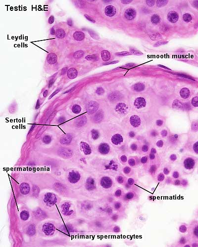
Sperm Under Microscope with Labeled Diagram » AnatomyLearner >> The ... Sperm under microscope 400x labeled. I will show you the sperm under a microscope 400x with the labeled diagram. Here in the diagram, you will see some seminiferous tubules lined by the thick germinal epithelium. The picture shows the dark Type A and pale Type B spermatogonia located at the seminiferous tubules' basal part.

Structure of Compact Bone
PDF Label parts of the Microscope: Answers Label parts of the Microscope: Answers Coarse Focus Fine Focus Eyepiece Arm Rack Stop Stage Clip . Created Date: 20150715115425Z ...
PID - Trimastix Ultrastructure
Label the Microscope Diagram | Download Scientific Diagram Download scientific diagram | Label the Microscope Diagram from publication: Laboratory Exercises in Microbiology: Discovering the Unseen World through Hands-on Investigation | Microbiology ...

astronomical optics, part 2: telescope & eyepiece combined
Blood Histology Slides with Description and Labeled Diagram The blood is a specialized connective tissue that is fluid and circulates through the vascular channel. In the blood histology slide, you will find different types of cells with their specific features. This might be a short article where I will show you all the cells from the blood microscope slide with a labeled diagram and actual pictures.
2011 Lab 1 - Spermatogenesis - Embryology
Labeling the Parts of the Microscope | Microscope activity, Science ... Jan 13, 2016 - Free worksheets for labeling parts of the microscope including a worksheet that is blank and one with answers. Pinterest. Today. Explore. ... Print a microscope diagram, microscope worksheet, or practice microscope quiz in order to learn all the parts of a microscope. CCabreza. Biology. Teaching Cells. Computer Science.

One Teacher's Adventures: Grade 8 Animal Cell Models
Labeling the Parts of the Microscope Labeling the Parts of the Microscope. This activity has been designed for use in homes and schools. Each microscope layout (both blank and the version with answers) are available as PDF downloads. You can view a more in-depth review of each part of the microscope here.
Quia - Protist Vocabulary
Microscope labeled diagram - SlideShare Microscope labeled diagram 1. The Microscope Image courtesy of: Microscopehelp.com Basic rules to using the microscope 1. You should always carry a microscope with two hands, one on the arm and the other under the base. 2. You should always start on the lowest power objective lens and should always leave the microscope on the low power lens ...

Vessel Comparison | BioNinja
Simple Microscope - Diagram (Parts labelled), Principle, Formula and Uses Simple microscope is a magnification apparatus that uses a combination of double convex lens to form an enlarged, erect image of a specimen. The working principle of a simple microscope is that when a lens is held close to the eye, a virtual, magnified and erect image of a specimen is formed at the least possible distance from which a human eye ...
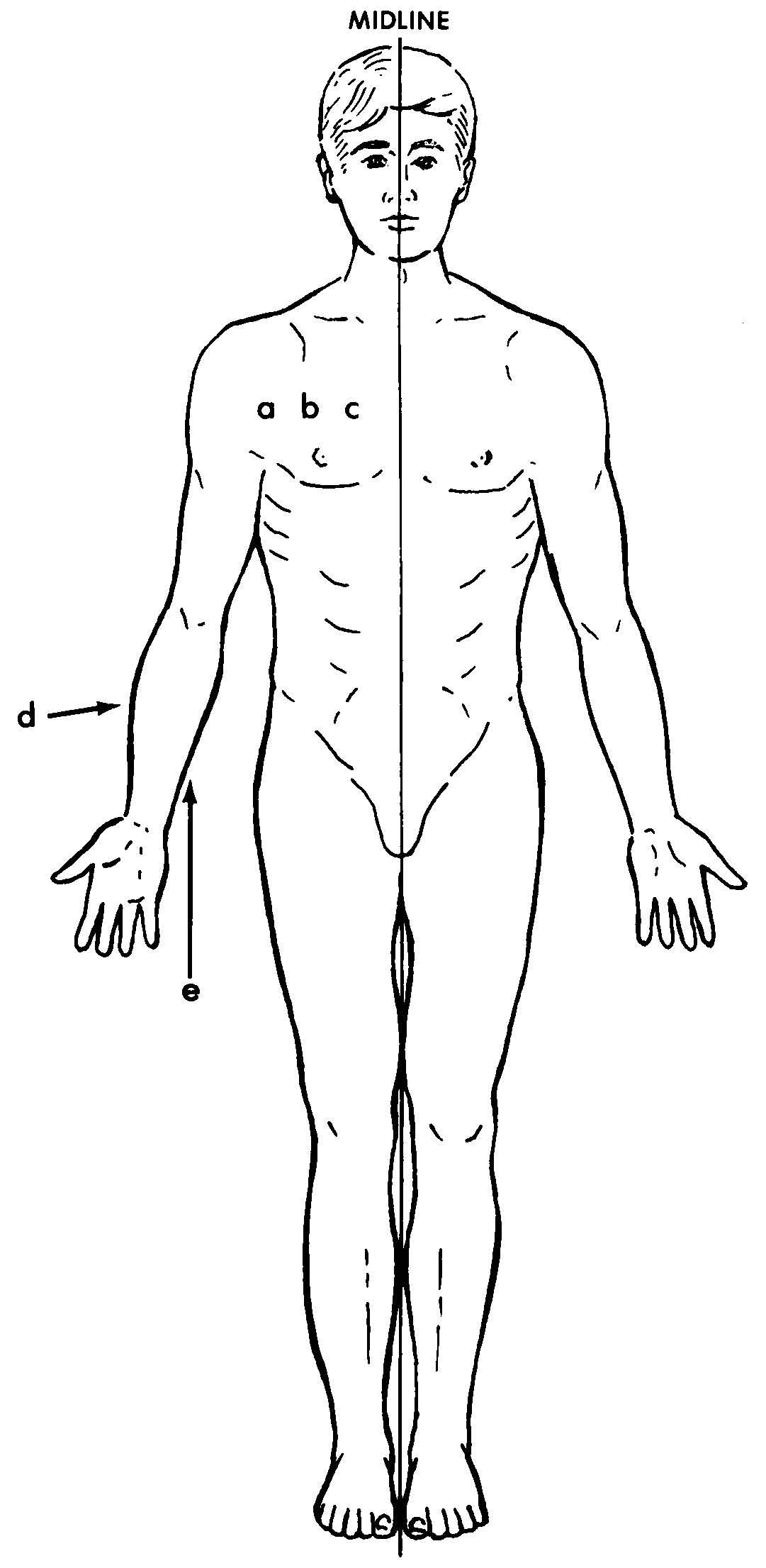
Images 01. Introduction and Terminology | Basic Human Anatomy
22 Parts Of a Microscope With Their Function And Labeled Diagram The field diaphragm control is located around the lens located in the base. Hinge Screw -This screw fixes the arm to the base and allow for the tilting of the arm. Stage Clips - They hold the slide firmly onto the stage. On/OFF Switch - This switch on the base of the microscope turns the illuminator off and on.
Post a Comment for "38 microscope diagram with labels"