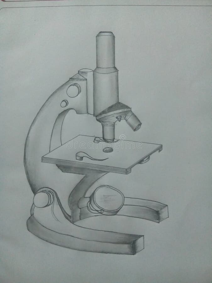
42 draw and label a compound microscope
Labelled Diagram of Compound Microscope - Biology Discussion The below mentioned article provides a labelled diagram of compound microscope. Part # 1. The Stand: The stand is made up of a heavy foot which carries a curved inclinable limb or arm bearing the body tube. The foot is generally horse shoe-shaped structure (Fig. 2) which rests on table top or any other surface on which the microscope in kept. Compound Microscope: Definition, Diagram, Parts, Uses, Working ... - BYJUS A compound microscope is defined as. A microscope with a high resolution and uses two sets of lenses providing a 2-dimensional image of the sample. The term compound refers to the usage of more than one lens in the microscope. Also, the compound microscope is one of the types of optical microscopes. The other type of optical microscope is a ...
Draw And Label A Microscope - Free PDF File Sharing Draw and label two or three Elodea cells, including cell wall, ... Observe the protists under the microscope. 4. Draw several protists and estimate their length. [Filename: cells_microscopy.pdf] - Read File Online - Report Abuse. ... Parts of the Light Microscope Compound Light Microscope Label each part and complete its description. T. Trimpe ...
Draw and label a compound microscope
The Compound Microscope.docx - The Compound Microscope 1. Draw and ... Draw and label a compound microscope. Parts FunctionsParts of a Compound Microscope 2. Enumerate all the parts of the microscope and give their corresponding functions. Tabulate your answer. a. Objective Lenses -forms the inverted image of the specimen and gives the initial magnification. -used for visualization of specimen. c. Parts of a Compound Microscope (And their Functions) List of Microscope Parts and their Functions. 1. Ocular Tubes (Monocular, Binocular & Trinocular) The ocular tubes, are to tubes that lead from the head of the microscope out to your eyes. On the end of the ocular tubes are usually interchangeable eyepieces (commonly 10X and 20X) that increase magnification. 16 Parts of a Compound Microscope: Diagrams and Video In compound microscopes with two eye pieces there are prisms contained in the body that will also split the beam of light to enable you to view the image through both eye pieces. 2. Arm. The arm of the microscope is another structural piece. The arm connects the base of the microscope to the head/body of the microscope.
Draw and label a compound microscope. Compound Microscope- Definition, Labeled Diagram, Principle, Parts, Uses Apr 03, 2022 · A compound microscope is of great use in pathology labs so as to identify diseases. Various crime cases are detected and solved by drawing out human cells and examining them under the microscope in forensic laboratories. The presence or absence of minerals and the presence of metals can be identified using compound microscopes. Draw a labelled diagram of an image formed by a compound microscope ... Draw a labelled diagram of an image formed by a compound microscope, with the image at least distance of distinct vision. Write any one expression for its magnifying power. Medium Solution Verified by Toppr Expression of magnifying power of a compound microscope is given by: m=− u ov o(1+ f eD) Microscope Activity - MICROBIOLOGY - 1... Draw a compound microscope. 2 ... A microscope is said to be parfocal when an ocular lens of a microscope don't lose focus when the objective lenses in use are adjusted while trying to find a better view for the sample. It is useful when the microscope have parfocal feature so the user or the phlebotomist don't need to adjust the focus when changing the power of ... How to draw compound of Microscope easily - step by step I will show you " How to draw compound of microscope easily - step by step "Please watch carefully and try this okay.Thanks for watching.....#microscopedrawi...
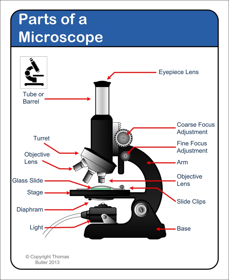
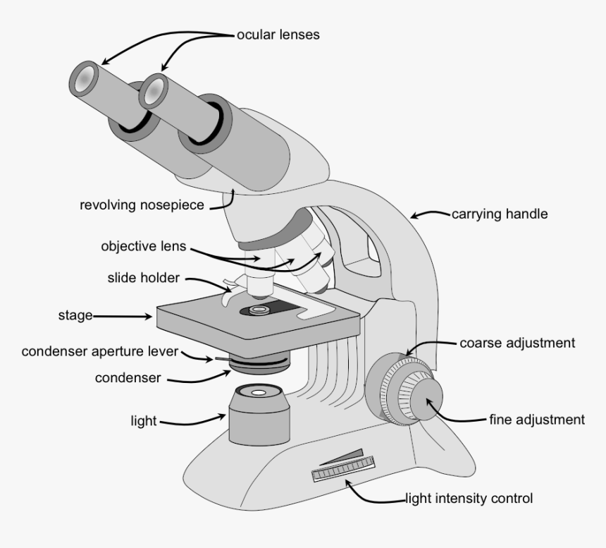
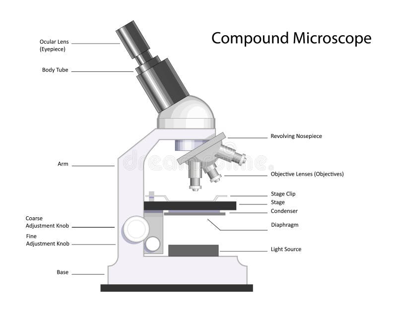
Compound Microscope: Parts of Compound Microscope - BYJUS (A) Mechanical Parts of a Compound Microscope 1. Foot or base It is a U-shaped structure and supports the entire weight of the compound microscope. 2. Pillar It is a vertical projection. This stands by resting on the base and supports the stage. 3. Arm The entire microscope is handled by a strong and curved structure known as the arm. 4. Stage Microscope Parts and Functions The specimen is placed on the glass and a cover slip is placed over the specimen. This allows the slide to be easily inserted or removed from the microscope. It also allows the specimen to be labeled, transported, and stored without damage. Stage: The flat platform where the slide is placed. Stage clips: Metal clips that hold the slide in place. Compound Microscope Parts - Labeled Diagram and their Functions - Rs ... Labeled diagram of a compound microscope Major structural parts of a compound microscope There are three major structural parts of a compound microscope. The head includes the upper part of the microscope, which houses the most critical optical components, and the eyepiece tube of the microscope. how to draw microscope (compound) - YouTube drawing microscope. Thank you watching more videos.please subscribe my channel
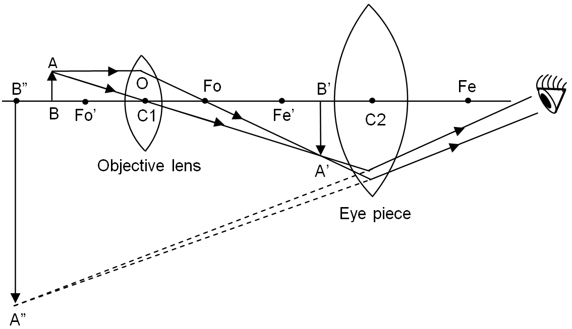
Compound Microscope - Types, Parts, Diagram, Functions and Uses A compound microscope has two convex lenses; an objective lens and eye piece. The objective lens is placed towards the object and the eyepiece is the lens towards our eye. Both eyepiece and objective lenses have a short focal length and fitted at the free ends of two sliding tubes. (4, 5, and 6) Compound microscope parts and magnification Draw a labelled ray diagram of a compound microscope and ... - Vedantu Answer. Verified. 123.4k + views. (Image 1 to be added soon) A tiny object AB to be magnified is placed in front of the objective lens just beyond its principal focus fo'. In this case, the objective lens O of the compound microscope forms a real, inverted and enlarged image A'B' of the object. Now A'B' acts as an object for the ... A Study of the Microscope and its Functions With a Labeled Diagram To better understand the structure and function of a microscope, we need to take a look at the labeled microscope diagrams of the compound and electron microscope. These diagrams clearly explain the functioning of the microscopes along with their respective parts. Man's curiosity has led to great inventions. The microscope is one of them. draw and label the compound microscope - Brainly.ph Draw and label the compound microscope - 9474237 samanthasolito19 samanthasolito19 19.01.2021 Science Elementary School answered Draw and label the compound microscope 1 See answer Advertisement Advertisement gemjem60 gemjem60 Answer: here I hope this will help.
Draw a labelled ray diagram of a compound microscope and ...
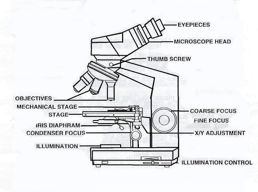
Parts of a microscope with functions and labeled diagram Head - This is also known as the body. It carries the optical parts in the upper part of the microscope. Base - It acts as microscopes support. It also carries microscopic illuminators. Arms - This is the part connecting the base and to the head and the eyepiece tube to the base of the microscope.
Compound Microscope Parts, Diagram Definition, Application ...
(b) Why both objective and eyepiece of a compound microscope must have ... (a) Draw the labelled ray diagram for the formation of image by a compound microscope. Derive an expression for its total magnification (or magnifying power), when the final image is formed at the near point. (b) Why both objective and eyepiece of a compound microscope must have short focal lengths?
Compound microscope stock illustration. Illustration of model ...
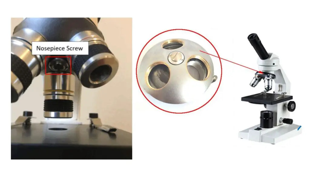
Compound Microscope - Diagram (Parts labelled), Principle and Uses Feb 03, 2022 · What are the 13 parts of a microscope? 1. Eyepiece 2. Eyepiece Tube 3. Objective Lens 4. Stage 5. Stage Clips 6. Nosepiece 7. Fine and Coarse Focus knobs 8. Illuminator 9. Aperture 10. Iris Diaphragm 11. Condenser 12. Condenser Focus Knob 13. The Rack stop Q 5. What are the 11 parts of a compound microscope?

Simple Microscope - Diagram (Parts labelled), Principle ...
Draw a neat labelled diagram of a compound microscope and ... - Sarthaks Dividing and multiplying by I1 G1 on the right side, we get Magnifying power of the objective (m0) = I1G1/OJ = Height of the image due to the objective. Magnifying power of the eye piece (me) = IG/I1G1 = Height of the final image / Height of the object for the eyepiece. ∴ m = m0 × me ..... (1)
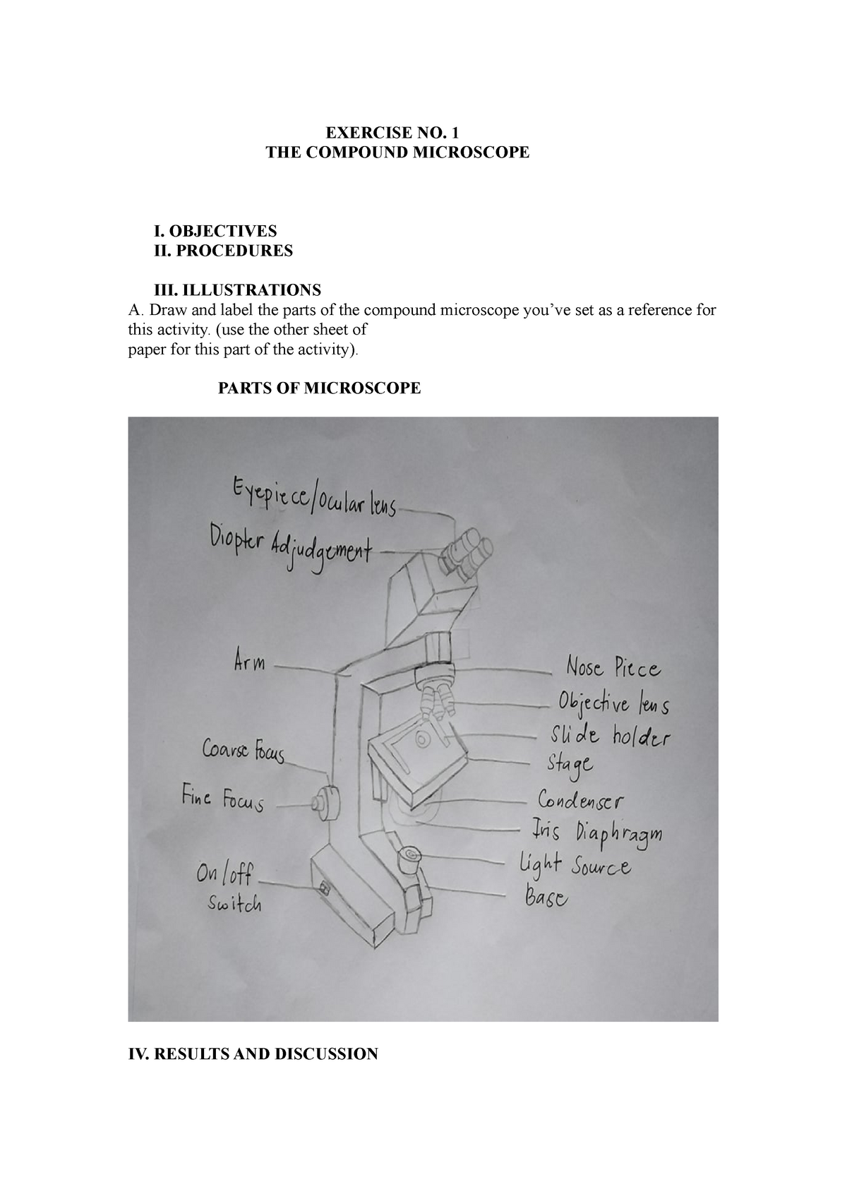
Compound microscopic microbiology - EXERCISE NO. 1 THE ...
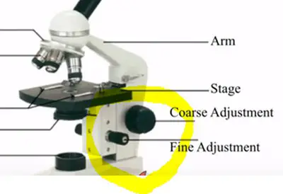
Parts of the Microscope with Labeling (also Free Printouts) 5. Knobs (fine and coarse) By adjusting the knob, you can adjust the focus of the microscope. The majority of the microscope models today have the knobs mounted on the same part of the device. Image 5: The circled parts of the microscope are the fine and coarse adjustment knobs. Picture Source: bp.blogspot.com.

Lasec Education | Key parts of a compound microscope and how ...
Diagram of a Compound Microscope - Biology Discussion 1. It is noted first that which objective lens is in use on the microscope. 2. Stage micrometer is positioned in such a way that it is in the field of view. 3. The eyepiece is rotated so that the two scales, the eyepiece or ocular scale and the stage micrometer scale, are parallel. 4.

Living Environment Course
(i) Draw a neat labelled ray diagram of a compound microscope. Explain ... The eyepiece forms its image A'' B'' which is virtual, erect and magnified. Thus the final image A'' B'' formed by the microscope is inverted and magnified and its position is outside the objective and eyepiece towards objective lens. Magnifying power of compound microscope is. for final image at distance of distinct vision. for final image at ...

Microscope Diagram Labeled, Unlabeled and Blank | Parts of a ...
PDF Draw And Label The Compound Microscope may 1st, 2019 - to draw an image formed by compound microscope first draw a straight horizontal line principle axis now make a lens take its focal length about 1 5cm at both sides now at one side draw an biology 1001 laboratory 4 microscopes measurements and may 12th, 2019 - 1 set up a compound microscope for use 2 answer the questions on the lab …

How to Draw a Microscope and Label Its Parts
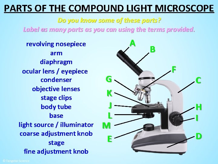
Label the microscope — Science Learning Hub Use this with the Microscope parts activity to help students identify and label the main parts of a microscope and then describe their functions. Drag and drop the text labels onto the microscope diagram. If you want to redo an answer, click on the box and the answer will go back to the top so you can move it to another box.

compound microscope Diagram | Quizlet
Compound Microscope Parts, Functions, and Labeled Diagram Nov 18, 2020 · Compound Microscope Definitions for Labels. Eyepiece (ocular lens) with or without Pointer: The part that is looked through at the top of the compound microscope. Eyepieces typically have a magnification between 5x & 30x. Monocular or Binocular Head: Structural support that holds & connects the eyepieces to the objective lenses.
Ilustrasi Stok Compound Microscope Line Drawing Structure ...
16 Parts of a Compound Microscope: Diagrams and Video In compound microscopes with two eye pieces there are prisms contained in the body that will also split the beam of light to enable you to view the image through both eye pieces. 2. Arm. The arm of the microscope is another structural piece. The arm connects the base of the microscope to the head/body of the microscope.

How to draw compound of Microscope easily - step by step
Parts of a Compound Microscope (And their Functions) List of Microscope Parts and their Functions. 1. Ocular Tubes (Monocular, Binocular & Trinocular) The ocular tubes, are to tubes that lead from the head of the microscope out to your eyes. On the end of the ocular tubes are usually interchangeable eyepieces (commonly 10X and 20X) that increase magnification.
Free Microscope Drawing, Download Free Microscope Drawing png ...
The Compound Microscope.docx - The Compound Microscope 1. Draw and ... Draw and label a compound microscope. Parts FunctionsParts of a Compound Microscope 2. Enumerate all the parts of the microscope and give their corresponding functions. Tabulate your answer. a. Objective Lenses -forms the inverted image of the specimen and gives the initial magnification. -used for visualization of specimen. c.
microscope drawing with label - Clip Art Library

Compound Microscope- Definition, Labeled Diagram, Principle ...

Microscopy
Parts of the Microscope with Labeling (also Free Printouts ...

How to Use a Compound Microscope: 11 Steps (with Pictures)

Can someone can send me diagram of this compound microscope ...
Parts Of A Microscope - Parts Of A Compound Microscope, HD ...

draw a microscope and level it's parts? - Brainly.in
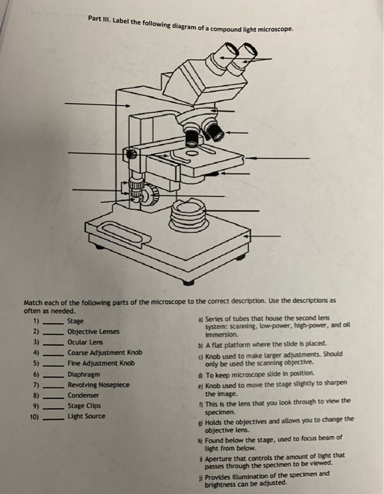
Solved Part III. Label the following diagram of a compound ...

The Compound Microscope parts & how they work
PRACTICAL BOOKLET - BIOLOGY4ISC

Microscope Drawing Worksheet | Clipart library - Free Clipart ...
Compound Microscope Parts, Diagram Definition, Application ...

Compound Microscope Parts – Labeled Diagram and their ...

Compound Microscope Parts, Functions, and Labeled Diagram ...
Compound Microscope stock vector. Illustration of research ...

Compound Microscope Parts, Functions, and Labeled Diagram ...

Difference between Simple and Compound Microscope ...
Compound Microscope - Types, Parts, Diagram, Functions and ...
i) Draw a neat labelled ray diagram of a compound microscope ...

How to draw Compound microscope | Microscope diagram easily Draw | SSC diagram

Labeling the Parts of the Microscope | Microscope World Resources

This is a common compound microscope. Label its parts from A ...

Compound Microscope Parts, Diagram Definition, Application ...

Compound Microscope

Diagram of a Compound Microscope

Microscope With Labels Clip Art at Clker.com - vector clip ...
MICROSCOPE PARTS PARTS OF THE COMPOUND LIGHT MICROSCOPE

File:Compound Microscope Drawing.jpg - Wikimedia Commons
Post a Comment for "42 draw and label a compound microscope"